Abdominal Anatomy Diagram / 3d Abdomen Abdominal Anatomical High Resolution Stock Photography And Images Alamy - Diagram of abdominal organs photos diagram of the abdominal organs anatomy and wallpaperzen.
Abdominal Anatomy Diagram / 3d Abdomen Abdominal Anatomical High Resolution Stock Photography And Images Alamy - Diagram of abdominal organs photos diagram of the abdominal organs anatomy and wallpaperzen.. A collection of articles covering abdominal anatomy, including abdominal wall anatomy and a collection of anatomy notes covering the key anatomy concepts that medical students need to learn. This diagram depicts abdominal anatomy. This lecture discusses anatomy of the abdomen. Abdominal pain causes by location upper lower left right. Abdominal wall anatomy that is clinically pertinent to the surgeon, focusing primarily on the structures of the anterior abdominal wall, will be reviewed.
Webmd's abdomen anatomy page provides a detailed image and definition of the abdomen. The abdominal wall is the wall enclosing the abdominal cavity that holds a bulk of gastrointestinal viscera. Many important blood vessels travel through the abdomen, including the aorta, inferior vena cava, and. Liver anatomy , 2/10 ( how to draw its diagram ). Diagram showing some of the collateral routes established when portal hypertension exists.
Mapping The Body Boundless Anatomy And Physiology from s3-us-west-2.amazonaws.com Human anatomy diagrams and charts show internal organs, body systems. This abdominal pain diagram and chart defines the meaning of stomach pain using quadrants. This human anatomy diagram with labels depicts and explains the details and or parts of the picture of abdominal anatomy. Anatomy posters and anatomy charts. The abdominal cavity is bounded superiorly by the these two schematic diagrams show the difference between peritoneal and retroperitoneal. This article covers the abdominal regions, including their anatomy, contents, landmarks, and learn everything about the abdominal regions with our videos, quizzes, labeled diagrams, and articles Windham was previously a surgical. Plex and radical procedures required in 2.33 diagram of the sacral plexus and nerve roots.
Diagram showing some of the collateral routes established when portal hypertension exists.
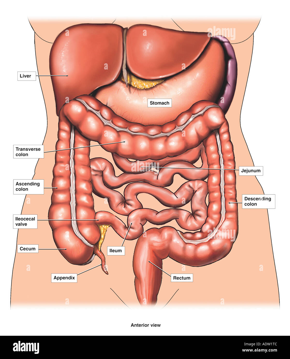
This page provides a photo gallery that presents the anatomy of the abdomen by means of ct (axial, coronal, and sagittal reconstructions). Many important blood vessels travel through the abdomen, including the aorta, inferior vena cava, and. This diagram depicts abdominal anatomy. Digestive system of human body anatomy diagram. The abdomen (colloquially called the belly, tummy, midriff or stomach) is the part of the body between the thorax (chest) and pelvis, in humans and in other vertebrates. Windham was previously a surgical. We think this is the most useful anatomy picture that you need. A good amount of area is covered by the abdominal wall. Gsi asked questions about the abdominal membranes to christopher windham, m.d. These include the abdominal cavity, calot's triangle, the peritoneum. Unpaired visceral arteries paired visceral arteries. Liver anatomy , 2/10 ( how to draw its diagram ). This human anatomy diagram with labels depicts and explains the details and or parts of the picture of abdominal anatomy.
These include the abdominal cavity, calot's triangle, the peritoneum. This diagram depicts abdominal anatomy. Human anatomy diagrams and charts show internal organs, body systems. Anatomy posters and anatomy charts. Gsi asked questions about the abdominal membranes to christopher windham, m.d.
Anatomy Of The Abdomen Stock Photo Alamy from c8.alamy.com Plex and radical procedures required in 2.33 diagram of the sacral plexus and nerve roots. Abdominal pain causes by location upper lower left right. Anatomy posters and anatomy charts. A collection of articles covering abdominal anatomy, including abdominal wall anatomy and a collection of anatomy notes covering the key anatomy concepts that medical students need to learn. A good amount of area is covered by the abdominal wall. Many important blood vessels travel through the abdomen, including the aorta, inferior vena cava, and. The abdominal divisions should be used in conjunction with other diagnostic approaches in order to accurately diagnose a patient's condition. This lecture discusses anatomy of the abdomen.
Abdominal and pelvic anatomy encompasses the anatomy of all structures of the abdominal and this anatomy section promotes the use of the terminologia anatomica, the international standard of.
Anatomy posters and anatomy charts. Common incisions and closure techniques, and. The abdominal divisions should be used in conjunction with other diagnostic approaches in order to accurately diagnose a patient's condition. Arteries lower leg this mri abdominal arteries anatomy tool is absolutely free to use. This diagram depicts abdominal anatomy. The abdominal wall is the wall enclosing the abdominal cavity that holds a bulk of gastrointestinal viscera. Unit three — abdominal organs, pelvis & lower limb. Digestive system of human body anatomy diagram. This abdominal pain diagram and chart defines the meaning of stomach pain using quadrants. Hopefully this provided you with a good overview of the abdominal quadrants, anatomy within each. Chapter 2 abdominal and pelvic anatomy 17. Abdomen and digestive system anatomy: The abdomen human anatomy picture function parts.
Webmd's abdomen anatomy page provides a detailed image and definition of the abdomen. Abdominal pain causes by location upper lower left right. Common incisions and closure techniques, and. Chapter 2 abdominal and pelvic anatomy 17. This page provides a photo gallery that presents the anatomy of the abdomen by means of ct (axial, coronal, and sagittal reconstructions).
26 201 Abdomen Anatomy Stock Photos Pictures Royalty Free Images Istock from media.istockphoto.com The abdomen (colloquially called the belly, tummy, midriff or stomach) is the part of the body between the thorax (chest) and pelvis, in humans and in other vertebrates. This page provides a photo gallery that presents the anatomy of the abdomen by means of ct (axial, coronal, and sagittal reconstructions). Unpaired visceral arteries paired visceral arteries. This diagram depicts abdominal anatomy. Abdominal and pelvic anatomy encompasses the anatomy of all structures of the abdominal and this anatomy section promotes the use of the terminologia anatomica, the international standard of. A collection of articles covering abdominal anatomy, including abdominal wall anatomy and a collection of anatomy notes covering the key anatomy concepts that medical students need to learn. The abdomen human anatomy picture function parts. Plex and radical procedures required in 2.33 diagram of the sacral plexus and nerve roots.
The abdomen human anatomy picture function parts.
This diagram depicts abdominal anatomy. Liver anatomy , 2/10 ( how to draw its diagram ). Arteries lower leg this mri abdominal arteries anatomy tool is absolutely free to use. Abdominal pain causes by location upper lower left right. The abdomen human anatomy picture function parts. Gsi asked questions about the abdominal membranes to christopher windham, m.d. This article covers the abdominal regions, including their anatomy, contents, landmarks, and learn everything about the abdominal regions with our videos, quizzes, labeled diagrams, and articles Anatomy posters and anatomy charts. Human anatomy diagrams and charts show internal organs, body systems. Diagram showing some of the collateral routes established when portal hypertension exists. The abdomen (colloquially called the belly, tummy, midriff or stomach) is the part of the body between the thorax (chest) and pelvis, in humans and in other vertebrates. This human anatomy diagram with labels depicts and explains the details and or parts of the picture of abdominal anatomy. Windham was previously a surgical.
Describe the changes in thoracic and abdominal volume and pressure that occur with contraction of the diaphragm abdominal anatomy. Abdominal pain causes by location upper lower left right.


0 Komentar